Big Bore RT
Big Bore RT
Big Bore RT
Big Bore RT
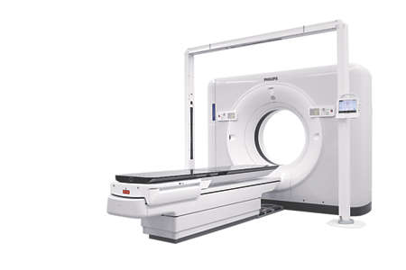
Big Bore RT is designed as a CT simulator to enhance clinical confidence, accelerate time to treat and maximize value of its investment without compromising on patient experience – four dimensions that are essential towards excellent care.
Clinical image gallery
- BBRT Abdomen and Pelvis
- BBRT Brain iDose IMR
- BBRT Abdomen for adenocarcinoma of the rectum
- BBRT Abdomen and Pelvis with O-MAR
- BBRT lung CT Full FOV 3mm slice thickness
- BBRT Abdomen 50 cm FOV and 2mm Slice thickness
- BBRT BI-lateral knee
- BBRT Chest abdomen pelvis
- BBRT Abdomen IMR
- BBRT Abdomen iDose IMR
- iDose⁴ with O-MAR


Features
Focusing on what matters in radiation oncology
Big Bore RT offers advanced tools to facilitate accurate efficient patient marking and simulation workflow. Featuring 60 cm true scan FOV for full anatomic visualization. It provides spatial positioning accuracy of less than 2mm between the imaging plane and the laser marking plane for confidence in patient marking. This meets the AAPM TG 66.

Where imaging and treatment planning meet
While excellent image quality is the foundation for accurate contouring and treatment plans, radiation oncology demands more. The Big Bore RT offers superb imaging and simulation solutions that integrate with your department workflow for the accuracy and efficiency cancer care requires.

Accelerate time to treatment
Efficiently move from scan to plan with virtual simulation and patient marking integrated at the scanner. Multimodality Simulation Workspace is a vendor-neutral image simulation platform that supports image fusion and contouring for all available images and data sets. It provides clinical teams with the tools necessary for multimodality image fusion, auto-contouring, and efficient collaborations helping to reduce patient wait time while providing quality care.

Specifications
- Features
- Bore size
- 85 cm
- Scan field of view
- 60 cm
- Gantry tilt
- -30° to +30° w/0.5° increments
- Generator power
- 80 kW
Related products
- IMR sets a new direction in CT image quality with industry-leading low-contrast resolution and virtually noise*-free images. Innovations in hardware and the reconstruction algorithm result in a reconstruction speed – less than three minutes for the majority of reference protocols – that allows model-based benefits to be achieved in even the most demanding applications.
- Designed to bring clarity and personalization in radiation oncology closer to the point of care, Philips Multimodality Simulation Workspace connects to Philips and non-Philips imaging devices or PACS to access image datasets including CT, MR, PET, and Spectral CT. It provides vendor-neutral support for multimodality image fusion and contouring to help physicians better define tumor volume and surrounding organs-at-risk.


Disclaimer
* In clinical practice, the use of IMR may reduce CT patient dose depending on the clinical task, patient size, anatomical location, and clinical practice. A consultation with a radiologist and a physicist should be made to determine the appropriate dose to obtain diagnostic image quality for the particular clinical task. Lower image noise, improved spatial resolution, improved low-contrast detectability, and/or dose reduction, were tested using reference body protocols. All metrics were tested on phantoms. Dose reduction assessments were performed using 0.8 mm slices, and tested on the MITA CT IQ Phantom (CCT183, The Phantom Laboratory), using human observers. Data on file.
** Image noise as defined by IEC standard 61223-3-5. Image noise was assessed using reference body protocol, on a phantom. Data on file